Super-resolution ultrasound localisation microscopy (SRUS/ULM)
Introduction
The spatial resolution of any imaging modality based on waves (electro-magnetic or mechanical) is fundamentally limited by the size of its wavelength (diffraction-limited image resolution). In biomedical ultrasound imaging this diffraction limit has resulted in an inherent compromise between spatial resolution and penetration depth. Recently such diffraction limit has been broken in the field of optical microscopy, by the so-called “super-resolved fluorescence microscopy” which was awarded the 2014 Novel Prize in Chemistry.
We are working on the ultrasound counterpart of this super resolution imaging, by developing techniques to detect and pin-point individual microbubble contrast agents within micro-vasculature. We have generated both in vitro and in vivo images of image resolution far below the diffraction limit. The figure below shows the first in-vivo super-resolved images of a mouse hear.
We are working with a number of collaborators to further improve the technique and apply this to a wide range of in vivo applications.
Please find below some further information on our technique and some current projects.
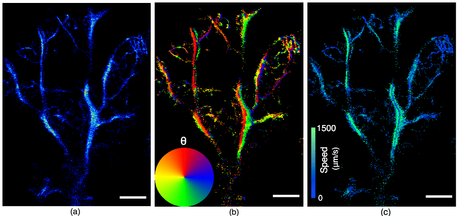
First in-vivo super-resolution images of a mouse hear. (a) Localisation Map. (b) Angle Map. (c) Velocity Map.
Super-resolution processing framework
We have developed a processing framework to localize and track high-concentration microbubbles at low frame rates, alleviating super-resolution imaging’s reliance on ultrafast ultrasound systems and long acquisition times that are often not available clinically. We have applied this framework to various imaging targets with different ultrasound systems. The effectiveness of the framework was validated by imaging animal brains using down sampled frame rates and human lymph nodes using data acquired by commercial systems in routine clinical management protocols.
The figures below show two recently reconstructed SR images of a human lymph node (left) and a rat brain (right) are shown below. Technical details can be found in. We are optimizing beamforming/reconstruction methods for 3D SR imaging and developing an easy-to-use interface for clinicians.

Super-resolution images of human lymph node (left) and a rat brain (right).
Current Projects
Find here all of our current projects around Super-Resolution :
- Project – 3D Super-Resolution Ultrasound Mouse Brain Vascular Atlas and Characterization.
- Project – Super resolution imaging of the myocardial microvasculature.
- Project – Super-resolution imaging of the gut microvasculature in rodents.
- Project – Coronary Ultrasound Localisation Microscopy.
- Project – Monitoring the breast tumour microenvironment following radiotherapy using super-resolution ultrasound and functional MRI.
- Project – Ultrafast 3-D Transcutaneous Super Resolution Ultrasound Using Row-Column Array Specific Coherence-Based Beamforming and Rolling Acoustic Sub-aperture Processing.
- Project – Non-invasive detection of lymph node metastasis.
- Project – Acoustic Wave Sparsely-Activated Localisation Microscopy (AWSALM).
- Project – Broad elevation project (BEP).
- Project – Enhancing super-resolution localisation ultrasound microscopy with model-based and data-driven approaches.
- Project – Image-based motion correction for cardiac imaging.
