Adaptive Weight-Based Beamforming
Project – Adaptive Weight-Based Beamforming
Quality of super-resolution imaging is significantly affected by microbubble isolation process. There is usually a trade-off between isolating too many side lobes and isolating too few main lobes for localisation, especially for 3D imaging with a matrix array where angle compounding cost much memory.
To deal with above-mentioned trade-off, we proposed to use beamformers based on adaptive weights for 3D super-resolution imaging, where only a single non-steering plane wave was transmitted from a matrix array. When conventional Delay and Sum Beamformer reconstructed a main lobe of weak signal weaker than a side lobe of strong signal in Field II simulation, the adaptive-weight-based beamformers changed the above intensity order, making it possible to isolate more main lobes in super-resolution processing. In vitro and in vivo experiments demonstrated that isolation with the adaptive beamforming contained more signals and less noise than that with DAS when the number of total isolations was similar.
Our proposed CV beamformer performed the best in all the comparisons. By using the CV beamformer and our bubble tracking technique, 3D super-resolution microflow was successfully reconstructed from a rabbit kidney within a 3-second acquisition.
The CV beamformer was also capable of being used with angle compounding, improving image quality in cardiovascular imaging with a phased array, as demonstrated in Extended Data Fig. 2 of our NBME paper.
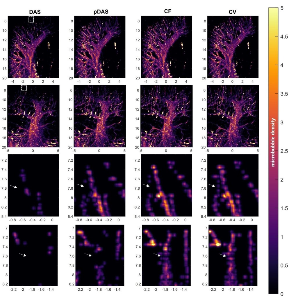
Maximum intensity projection of SRUS images of rabbit kidney. The four columns correspond to the DAS, p-DAS, CF, and CV beamformers, from left to right. First row: lateral-depth-plane MIPs; Second row: elevation-depth-plane MIPs; Third and fourth rows: Zoomed-in images of the area in the boxes shown in the first and second rows respectively. p-DAS: DAS with p-th root compression. CF: Coherence Factor. CV: Coherence Energy to Variance.
